

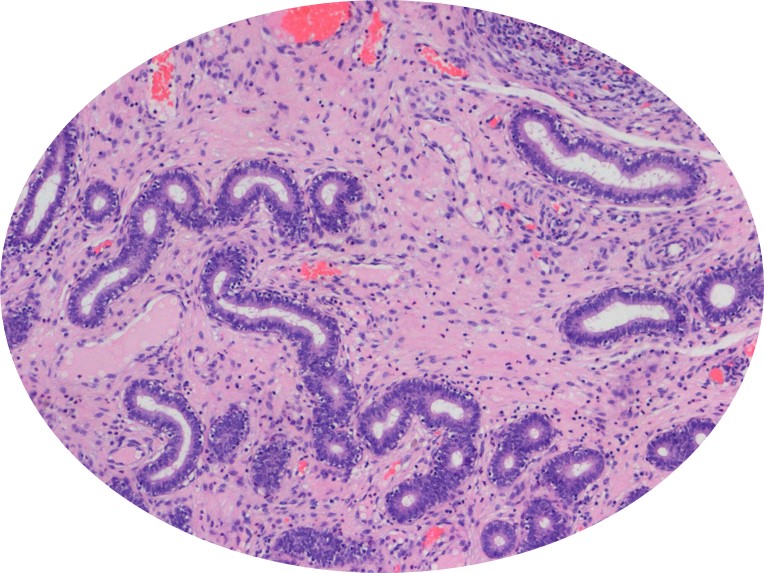
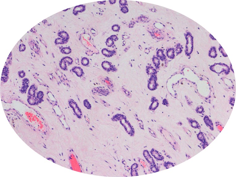
There are only a handful cases where a tissue, observed from outside, reveals details of its inner morphology. The Uterus is a prominent example of such a case.
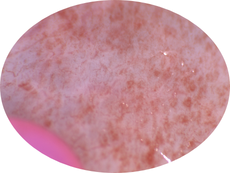
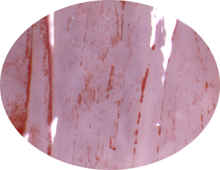
The rapid evolution of the uterus outermost layer – the endometrium – is clearly reflected on its outermost layer that is facing the uterus lumen.
Even a non-expert eye can easily distinguish between different phases of the uterus evolution.
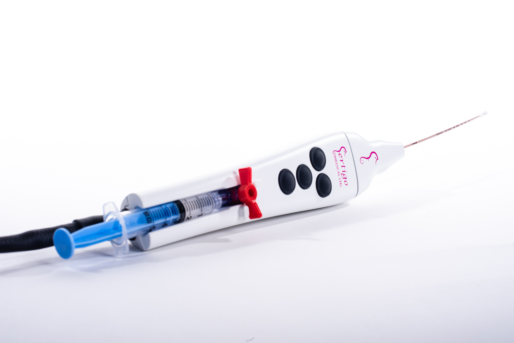
However, when it comes to quantification and precise determination of the day in the menstrual cycle, a scientific approach is in place. Fertigo Medical invented a device and method to discern the endometrium dating.
Special image acquisition and other parameters, combined with sophisticated image processing, has been pre-clinically proven to provide as good an estimate of the uterus status as do the traditional methods (e.g., histology, hormone level, estrus).